37 results filtered with: Tissue
- Digital Images
- Online
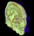
Structure of human head and brain
Parashkev Nachev
- Digital Images
- Online
Human heart (aortic valve) tissue displaying calcification
Sergio Bertazzo, Department of Materials, Imperial College London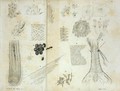
- Digital Images
- Online
Portrait of Samuel Thomas von Sommering [1755 - 1830], physician, anatomist, anthropologist, paleontologist and inventor, who discovered the macula in the retina of the human eye.
- Digital Images
- Online
Acute nephritis in calf kidneys
Michael Frank, Royal Veterinary College
- Digital Images
- Online
Acinar tissue - The flowers of diabetes
Odra Noel
- Digital Images
- Online
Human heart (mitral valve) tissue displaying calcification
Sergio Bertazzo, Department of Materials, Imperial College London
- Digital Images
- Online
Human heart (mitral valve) tissue displaying calcification
Sergio Bertazzo, Department of Materials, Imperial College London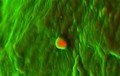
- Digital Images
- Online
Human heart (aortic valve) tissue displaying calcification
Sergio Bertazzo, Department of Materials, Imperial College London
- Digital Images
- Online
Tongue, squames on fungiform papilla, SEM
David Gregory & Debbie Marshall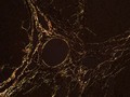
- Digital Images
- Online
Liver of a DEN (Diethylnitrosamine)-treated rat. DEN is a toxic chemical which quickly induces liver cirrhosis followed by HCC (Hepatocellular carcinoma, a primary liver cancer). Cirrhosis is an end result of fibrosis, the scarring of liver tissue. Fibrosis is caused by the overproduction of collagen, a component of the connective tissue forming the liver. To grade the amount of cirrhosis present in a liver sample, collagen is made visible using the dye sirius red. Under polarized light, collagen is observed as the golden to red color as shown in this image.
Tabea Hohensee
- Digital Images
- Online
Adipose Tissue
Odra Noel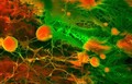
- Digital Images
- Online
Human heart (coronary artery) tissue displaying calcification
Sergio Bertazzo, Department of Materials, Imperial College London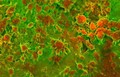
- Digital Images
- Online
Human heart (aorta) tissue displaying calcification
Sergio Bertazzo, Department of Materials, Imperial College London
- Digital Images
- Online
Human heart (mitral valve) tissue displaying calcification
Sergio Bertazzo, Department of Materials, Imperial College London
- Digital Images
- Online
Organic forms
Paul Griggs
- Digital Images
- Online
Human heart (aortic valve) tissue displaying calcification
Sergio Bertazzo, Department of Materials, Imperial College London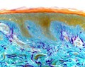
- Digital Images
- Online
Light micrograph of section of skin
Kevin Mackenzie, University of Aberdeen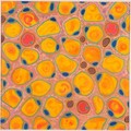
- Digital Images
- Online
'More to love' Adipose tissue
Odra Noel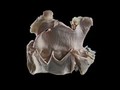
- Digital Images
- Online
Aortic valvular endocarditis, horse. This equine (horse) aorta has been dissected at the level of the valve, separating the artery from the left ventricle (bottom chamber). Nodules are present which have formed as a consequence of endocarditis.
Michael Frank, Royal Veterinary College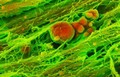
- Digital Images
- Online
Human heart (aortic valve) tissue displaying calcification
Sergio Bertazzo, Department of Materials, Imperial College London
- Digital Images
- Online
Structure of tissue of esophagus, stomach and intestines.
Emile Beau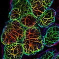
- Digital Images
- Online
3D depth-coloured transparent mouse mammary gland
Felicity Davis, Bethan Lloyd-Lewis and Christine Watson; University of Cambridge
- Digital Images
- Online
Structure of human head and brain
Parashkev Nachev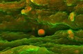
- Digital Images
- Online
Human heart (aortic valve) tissue displaying calcification
Sergio Bertazzo, Department of Materials, Imperial College London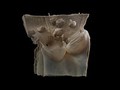
- Digital Images
- Online
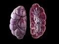
Arterial nodules, horse
Michael Frank, Royal Veterinary College