Three demonstrations on malformations of the hind end of the body / by Arthur Keith.
- Keith, Arthur, Sir, 1866-1955.
- Date:
- 1908
Licence: In copyright
Credit: Three demonstrations on malformations of the hind end of the body / by Arthur Keith. Source: Wellcome Collection.
Provider: This material has been provided by The Royal College of Surgeons of England. The original may be consulted at The Royal College of Surgeons of England.
5/42 (page 3)
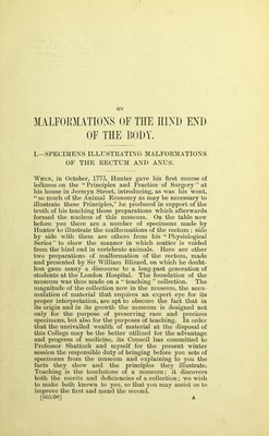
![Group II represents a greater degree of arrest of develop- ment, or of atrophy, of tlie rectum than is shown in Group I. Table I.—Specimens of Malformation of the JRectnm in London Museums. Groups. In Museum of Eoyal College of Surgeons. In Museums of Metro- ])olitau Medical Schools. Total A. Males (see Fig. 2): 1. Eectum opening in urethra 7 26 33 2. Eectum ending as cord at or above base of prostate 0 7 7 3. Eectum ending as cord at site of proctodaeum 2 5 7 4. Eectum ending blindly at procto- daeum 1 6 7 B._Pemales (see Fig. 4): 1. Eectum ending in vulva or vagina 1 5 6 2. Eectum ending in cord above upper fornix of vagina 0 5 5 3. Eectum ending as cord at upper fornix of vagina 0 3 3 4. Eectum ending as cord on vagina below upper fornix 0 2 2 5. Eectum ending blindly or as cord at site of proctodaeum 3 7 10 C. Miscellaneous Specimens : 1. Imperfect 1 5 G 2. Imperforate rectum in females with male form of external genital organs 3 3 6 3. Eectum opening abnormally ... 1 1 4. Eare malformations 2 3 5 5. Abnormalities of the rectum in domesticated animals IG 0 IG 37 77 114 Geoup III.—Males in ivliich the Bectum terminates in a Fibrous Cord at the Site of the Proctodaeum (Fig. 2). Of this group there are two examples in the College collection, and five in the school museums. The nature of this malformation and its relation to the other groups will be apparent when the embryology and evolution of the rectum is dealt with. A 2](https://iiif.wellcomecollection.org/image/b22472873_0007.jp2/full/800%2C/0/default.jpg)