Anatomy, descriptive and surgical / Edited by T. Pickering Pick and Robert Howden.
- Gray, Henry, 1827-1861.
- Date:
- 1901
Licence: Public Domain Mark
Credit: Anatomy, descriptive and surgical / Edited by T. Pickering Pick and Robert Howden. Source: Wellcome Collection.
Provider: This material has been provided by the Augustus C. Long Health Sciences Library at Columbia University and Columbia University Libraries/Information Services, through the Medical Heritage Library. The original may be consulted at the the Augustus C. Long Health Sciences Library at Columbia University and Columbia University.
173/1252 (page 177)
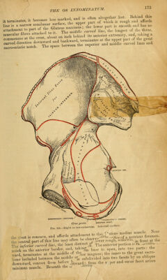
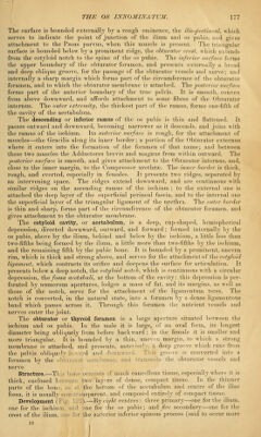
![The surface is bounded externally by a rough eminence, the ilio-peetineal, which serves to indicate the point of junction of the ilium and os pubis, and gives attachment to the Psoas parvus, when this muscle is present. The triangular surface is bounded below by a prominent ridge, the obturator crest, which extends from the cotyloid notch to the spine of the os pubis. The inferior surface forms the upper boundary of the obturator foramen, and presents externally a broad and deep oblique groove, for the passage of the obturator vessels and nerve; and internally a sharp margin which forms part of the circumference of the obturator foramen, and to which the obturator membrane is attached. The posterior surface forms part of the anterior boundary of the true pelvis. It is smooth, convex from above downward, and affords attachment to some fibres of the Obturator internus. The outer extremity, the thickest part of the ramus, forms one-fifth of the cavity of the acetabulum. The descending or inferior ramus of the os pubis is thin and flattened. It passes outward and downward, becoming narrower as it descends, and joins with the ramus of the ischium. Its anterior surface is rough, for the attachment of muscles—the Gracilis along its inner border; a portion of the Obturator externus where it enters into the formation of the foramen of that name; and between these two muscles the Adductores brevis and magnus from Avithin outward. The posterior surface is smooth, and gives attachment to the Obturator internus, and, close to the inner margin, to the Compressor urethra. The inner border is thick, rough, and everted, especially in females. It presents two ridges, separated by an intervening space. The ridges extend downward, and are continuous with similar ridges on the ascending ramus of the ischium ; to the external one is attached the deep layer of the superficial perineal fascia, and to the internal one the superficial layer of the triangular ligament of the urethra. The outer border is thin and sharp, forms part of the circumference of the obturator foramen, and gives attachment to the obturator membrane. The cotyloid cavity, or acetabulum, is a deep, cup-shaped, hemispherical depression, directed downward, outward, and forward; formed internally by the os pubis, above by the ilium, behind and below by the ischium, a little less than two-fifths being formed by the ilium, a little more than two-fifths by the ischium, and the remaining fifth by the pubic bone. It is bounded by a prominent, uneven rim, which is thick and strong above, and serves for the attachment of the cotyloid ligament, which contracts its orifice and deepens the surface for articulation. It presents below a deep notch, the cotyloid notch, which is continuous with a circular depression, the fossa acetabuli, at the bottom of the cavity: this depression is per- forated by numerous apertures, lodges a mass of fat, and its margins, as well as those of the notch, serve for the attachment of the ligamentum teres. The notch is converted, in the natural state, into a foramen by a dense ligamentous band which passes across it. Through this foramen the nutrient vessels and nerves enter the joint. The obturator or thyroid foramen is a large aperture situated between the ischium and os pubis. In the male it is large, of an oval form, its longest diameter being obliquely from before backward ; in the female it is smaller and more triangular. It is bounded by a thin, uneven margin, to which a strong membrane is attached, and presents, anterior] a deep groove which runs from the pelvis obliquely ii -d and d Th. I groove is converted into a foramen by the obi in I its the obturator vessels and nerve. Structure.—Tl e consists of much cancellous tissue, especially where it is thick, enclosed between lw« layers of dense, compact tissue. In the thinner parts of the bon the bottom of the acetabulum and centre of the iliac- fossa, it is usually isparent, and composed entirely of compact tissue. Development ( ).—By eight centres: three primary—one for the ilium, one for the ischium, md one for the os pubis; and five secondary—one for the crest of the ilium, one for the anterior inferior spinous process (said to occur more 12](https://iiif.wellcomecollection.org/image/b21220700_0173.jp2/full/800%2C/0/default.jpg)